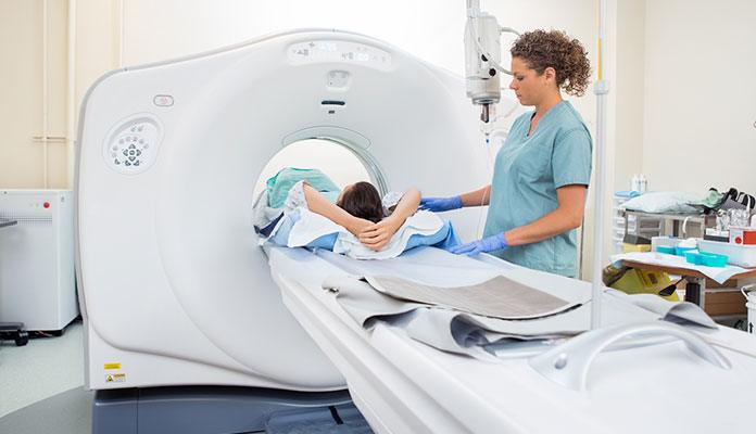
Computerized tomography (CT) scan

A CT scan involves combining a lot of X-ray images observed from different angles around the body and using computer processing to make a cross-sectional image that resembles slices of the chest. These images are usually detailed and show the state of the pleura and if there are other factors responsible for pain such as clotting of blood in the lungs. CT scans are done for further investigation if the normal chest X-ray doesn’t provide enough information. It allows the doctor to get a closer look at inflamed tissues.
Ultrasound
This is an imaging method that uses high-frequency sound waves to make exact images of structures in the body. Doctors use ultrasounds to observe the condition of the lungs and see if you have pleural effusion. Doctors are also able to see if there is any sign of inflammation in the lungs through an ultrasound.
Electrocardiogram (ECG or EKG)
Doctors recommend ECG to monitor heart activities. This is done to be sure there are no heart complications.


